


Find Your Next Running Race
Ready to put your training to the test? Here are some upcoming running events matched to this article.
 Coach Graeme

Last updated:
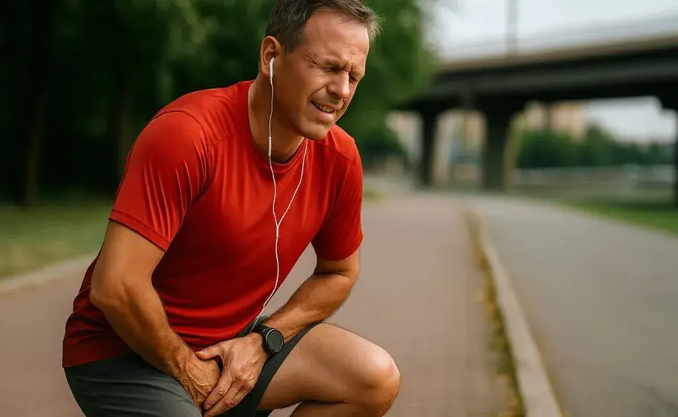
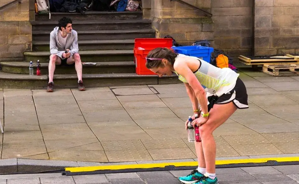
The calcaneus is the heel bone — the largest bone in the foot and the one that absorbs the full impact of each footstrike. At approximately 2–3 times body weight per stride, a runner covering 40km per week subjects the calcaneus to millions of repetitive loading cycles annually. When pain develops in or around the heel bone after running, it can have four quite different causes — each with a different location, different underlying mechanism, and a different treatment approach.
The most common mistake runners make with calcaneus pain is treating it generically: resting for a week, resuming training, and finding the pain returns immediately. Without identifying the specific cause, treatment tends to miss the mechanism. This guide covers how to distinguish the four main causes of calcaneus pain in runners, the red flags that require immediate cessation and clinical assessment, and the evidence-based treatment for each condition.
Not sure where to start with training?
Tell us your goal and schedule, and we’ll give you clear direction.
No obligation. Quick, practical advice.
Explore our running training articles for more helpful articles and resources.
The calcaneus forms the posterior (rear) section of the foot and is the foundation of the longitudinal arch. Several critical structures attach to it or pass adjacent to it: the Achilles tendon inserts at the posterior calcaneal tuberosity; the plantar fascia originates at the medial calcaneal tubercle on the underside; the retrocalcaneal bursa sits between the Achilles tendon and the posterior-superior surface; and a network of blood vessels, nerves, and ligaments traverse the surrounding soft tissue.
This anatomical complexity means that “calcaneus pain” is a location, not a diagnosis. Pain in the same general heel region can originate from any of these structures — and the distinction matters enormously for treatment. Treating plantar fasciitis protocols on what is actually an insertional Achilles tendinopathy, for example, can worsen the condition. Running through what is actually a calcaneal stress fracture risks a complete fracture requiring surgery.
| Pain location | Most likely diagnosis | Key distinguishing feature | Run through it? |
|---|---|---|---|
| Underside of heel (plantar surface) | Plantar fasciitis | Worst first steps in morning; improves with movement; worsens after long sitting | Reduced load, yes |
| Back of heel (Achilles insertion point) | Insertional Achilles tendinopathy | Worsens uphill; tender at bone-tendon junction; worsens in flat/minimal shoes | Reduced load, yes |
| Back and top of heel (posterior-superior) | Retrocalcaneal bursitis / Haglund's deformity | Aggravated by shoe collar; visible/palpable bump; improves in open-back shoes | Footwear modification, yes |
| Diffuse heel bone (medial, lateral, plantar) | Calcaneal stress fracture | Positive squeeze test; progressive during run; does not improve with warm-up | No — stop immediately |
Plantar fasciitis accounts for approximately 10% of all running-related injuries, making it the most prevalent cause of heel pain in runners. The plantar fascia is a thick band of connective tissue running from the medial calcaneal tubercle (the underside-front of the heel bone) to the bases of the toes, forming the primary support structure of the longitudinal arch. When repeatedly stressed beyond its tolerance — by overtraining, sudden mileage increases, poor footwear, or tight calf muscles — the tissue at its calcaneal origin becomes inflamed and degenerative.
The hallmark symptom is pain on the first steps in the morning or after a period of inactivity: the plantar fascia shortens during rest and is suddenly loaded when the foot hits the ground. This “post-static dyskinesia” typically improves after a few minutes of walking as the tissue warms and elongates, then worsens again after prolonged running or standing. Pain is located at the underside of the heel, slightly toward the inside of the foot, and is often very specific to direct pressure at that point.
Treatment: The most effective conservative management combines calf and plantar fascia stretching (particularly the first-thing-in-the-morning towel stretch before taking any steps), eccentric and isometric calf strengthening to reduce the load on the plantar fascia’s calcaneal insertion, and temporary reduction in running volume and intensity. Heel cups or semi-rigid orthotics can offload the insertion during the acute phase. Shoes with a moderate heel-to-toe drop (8–12mm) are generally preferable to flat or zero-drop shoes during recovery, as they reduce stretch on the plantar fascia at push-off. For chronic plantar fasciitis (present more than 3 months), extracorporeal shockwave therapy has the strongest evidence base among procedural interventions, with multiple RCTs demonstrating significant pain reduction at 3–6 months.
Insertional Achilles tendinopathy affects the bottom 2cm of the Achilles tendon where it attaches to the posterior calcaneus — distinct from mid-portion Achilles tendinopathy (which occurs 2–6cm above the insertion) and requiring different treatment. The pain is located at the back of the heel, directly at the bone-tendon junction, and is characteristically aggravated by uphill running, flat or minimal shoes, and any loading that increases the stretch on the Achilles insertion.
The critical distinction for treatment: the standard eccentric calf drop exercise (lowering the heel below the level of a step) — which is highly effective for mid-portion Achilles tendinopathy — is contraindicated for insertional Achilles tendinopathy. The increased stretch on the Achilles insertion from dropping below neutral compresses the tendon against the bone and aggravates the condition. Treatment instead uses isometric loading holds (standing calf raise held at the top position for 45–60 seconds, 4–5 times, performed multiple times per day) and concentric calf raises through a limited range (heel to neutral, not below), progressing to heavy slow resistance training over 8–12 weeks.
A small heel raise (6–8mm) inside the running shoe reduces the stretch on the Achilles insertion during running and typically produces immediate symptomatic relief, though it should be used as a temporary adjunct rather than a permanent solution. Avoiding barefoot walking, particularly in the morning, is also important during the acute phase. Uphill running and speed work should be eliminated initially and reintroduced only once pain during flat running has resolved.
Between the Achilles tendon and the posterior-superior surface of the calcaneus sits a small bursa — a fluid-filled sac that reduces friction between these structures. When the posterior heel is repeatedly compressed or rubbed — by the shoe counter digging into the area, by running in shoes with a rigid heel collar, or by the structural prominence that develops in Haglund’s deformity — this bursa becomes inflamed. The pain sits at the back and top of the heel, not at the Achilles insertion point lower on the bone or at the plantar surface, and is distinctly aggravated by shoe collar contact.
Haglund’s deformity is an abnormal bony prominence on the posterior-superior calcaneus that can be palpated as a hard bump at the back of the heel bone. It develops over time in runners who repeatedly stress this area and is a structural risk factor for retrocalcaneal bursitis. The “pump bump” — a related but slightly different bony prominence on the posterolateral calcaneus — produces similar symptoms and is named for its association with stiff-heeled footwear.
Treatment: The most immediately effective intervention is removing the source of compression — switching to open-back shoes or running shoes with a softer, more accommodating heel counter. A silicone heel pad that lifts the calcaneus away from the shoe collar can also help. Load reduction (reducing speed work and total weekly mileage) allows the bursae to settle. Ice applied directly to the posterior heel for 15–20 minutes after running reduces acute inflammation. For chronic or structurally driven cases, shockwave therapy or, rarely, surgical removal of the bony prominence is considered.
A calcaneal stress fracture is a partial crack in the heel bone caused by the cumulative effect of repeated impact loading — typically following a sudden, significant increase in training volume or intensity, a return to running after a prolonged break, or a period of running on particularly hard surfaces with inadequate footwear. It is the most serious of the four conditions and is frequently misdiagnosed as plantar fasciitis in the early stages, because the pain can initially feel similar and is located in the same general area.
The squeeze test: Apply firm pressure to both the medial (inside) and lateral (outside) of the calcaneus simultaneously, squeezing from side to side. In a calcaneal stress fracture, this medial-lateral compression reproduces the pain distinctly. This test is negative in plantar fasciitis and Achilles tendinopathy. A positive squeeze test in a runner with heel pain warrants immediate cessation of running and imaging. Note: A clinician should perform a definitive assessment — this is a screening tool, not a diagnostic replacement.
The distinguishing pattern of a stress fracture is progressive worsening: the pain increases as the run continues rather than easing after the first few minutes of warm-up. There is often diffuse tenderness over the entire heel bone rather than a precise point of maximal tenderness, and pain may persist at rest or at night. Standard X-rays are frequently negative in the early stages of calcaneal stress fractures — the fracture line may not be visible until 2–3 weeks after onset. MRI is the most sensitive imaging modality and can detect bone marrow oedema (the precursor to visible fracture) before the fracture is radiographically apparent. A PMC review of heel pain in athletes notes that calcaneal stress fractures are often associated with gait alterations and can precede plantar fascia ruptures if not identified and managed.
Treatment: Immediate cessation of all running and impact loading. Depending on severity, a walking boot or short-term non-weight-bearing may be required. Return to running typically begins no earlier than 6–8 weeks after diagnosis, following a progressive loading protocol. Bone health should be assessed — low bone density (from inadequate calcium/vitamin D, low energy availability, or the female athlete triad) is a risk factor for stress fractures in runners. Our shin splint exercises guide covers the related topic of tibial stress reactions and the training load principles that apply equally to calcaneal stress fractures.
Several training and biomechanical factors increase the risk of all calcaneus-related pain in runners. The most consistently identified is a rapid increase in training load — the 10% weekly mileage rule exists specifically because the calcaneus and its attached structures adapt more slowly to impact loading than cardiovascular fitness does. A runner whose aerobic system can handle 80km per week may have musculoskeletal structures that can only safely sustain 50km.
Calf and Achilles tightness increases the load transferred to the calcaneus at every footstrike. Tight calves increase the tension in the plantar fascia (which shares mechanical load with the gastrocnemius-soleus through their shared calcaneal attachment), and reduce ankle dorsiflexion range, forcing compensatory loading patterns. Regular calf stretching — gastrocnemius with the knee straight, soleus with the knee bent — and eccentric calf strengthening are the most evidence-supported interventions for reducing calcaneal loading across all four conditions. Our tibialis anterior exercises guide covers complementary lower leg strengthening that builds overall foot and ankle resilience.
Footwear is a modifiable risk factor. Running shoes should be replaced after approximately 600–800km of use — the midsole loses cushioning properties well before the outsole shows visible wear, meaning shoes that look fine may no longer be providing adequate impact attenuation. The heel-to-toe drop of the shoe matters most for insertional Achilles tendinopathy (higher drop = less Achilles stretch) and is less critical for plantar fasciitis and stress fractures where total cushioning and fit are more relevant.
Running cadence also influences calcaneal loading. A lower cadence (fewer steps per minute) produces a longer stride, more overstriding, and higher peak braking forces at heel contact. Increasing cadence by 5–10% — achievable within 4–6 weeks of deliberate practice — has been shown to reduce peak tibial acceleration and ground reaction forces, which translates to reduced load on the heel. Our guide on running with sore legs covers the broader training load management decisions that apply during recovery from heel injuries.
Many runners self-manage early-stage plantar fasciitis and Achilles tendinopathy successfully with load reduction, stretching, and appropriate footwear. However, certain presentations warrant prompt clinical assessment: any heel pain that does not improve within 2–3 weeks of conservative management; pain that is worsening rather than improving; a positive squeeze test; pain at rest or at night; heel pain in a runner who has recently significantly increased mileage (stress fracture risk); and bilateral heel pain (which may indicate a systemic cause).
A physiotherapist, sports medicine physician, or podiatrist can provide gait analysis, confirm the diagnosis, and prescribe the specific loading programme for each condition. For calcaneal stress fracture, imaging referral is required. For chronic plantar fasciitis and insertional Achilles tendinopathy, shockwave therapy has the strongest procedural evidence base and is available through sports physiotherapy clinics and sports medicine practices.
Train Smarter and Reduce Injury Risk
SportCoaching's running training plans and coaching build load progressively to stay within the musculoskeletal tolerance that prevents heel pain and overuse injuries — with specific guidance on when to modify, when to rest, and how to return to full training after an injury setback.
What causes calcaneus pain after running?
The four main causes are plantar fasciitis (underside of heel, worst in the morning), insertional Achilles tendinopathy (back of heel at tendon attachment), retrocalcaneal bursitis/Haglund’s deformity (posterior-superior heel, aggravated by shoe collar), and calcaneal stress fracture (diffuse bone pain that worsens during running). Each has a different location and pattern — identifying which is the first step to effective treatment.
How do I know if I have a calcaneal stress fracture?
The medial-lateral squeeze test is the most useful screening tool: firm simultaneous compression of both sides of the heel bone reproduces sharp pain in a stress fracture but not in plantar fasciitis or tendinopathy. Other signs: pain that worsens progressively during a run, pain at rest or at night, and no response to standard plantar fasciitis treatment. Stop running and seek imaging — X-ray is often negative early; MRI is more sensitive.
What is the difference between plantar fasciitis and insertional Achilles tendinopathy?
Plantar fasciitis: underside of heel, worst first steps in the morning, improves with movement. Insertional Achilles tendinopathy: back of heel at the tendon-bone junction, worsens uphill and in flat shoes. Treatment also differs critically: the eccentric heel drop exercise effective for mid-portion Achilles tendinopathy is contraindicated for the insertional variant.
Can I keep running with calcaneus pain?
Mild plantar fasciitis and early insertional Achilles tendinopathy: reduce load but continue running. Retrocalcaneal bursitis: footwear modification usually allows continued running. Calcaneal stress fracture: stop immediately. The test: if pain worsens during the run rather than easing after warm-up, stop and seek assessment.
How long does calcaneus pain after running take to heal?
Mild plantar fasciitis: 4–8 weeks. Chronic plantar fasciitis: 3–6 months. Insertional Achilles tendinopathy: 8–12 weeks. Calcaneal stress fracture: 10–14 weeks minimum (6–8 weeks immobilisation + gradual return). Retrocalcaneal bursitis: 4–6 weeks. All recover faster with early identification and appropriate management.
Ready to put your training to the test? Here are some upcoming running events matched to this article.


Head Coach & Founder, SportCoaching
Graeme is the founder of SportCoaching and has coached more than 750 athletes from 20 countries, from beginners to Olympians, in cycling, running, triathlon, mountain biking, boxing, and skiing. His coaching philosophy and methods form the foundation of SportCoaching's training programs and resources.
No matter your goals, SportCoaching offers tailored training plans to suit your needs. Whether you’re preparing for a race, tackling long distances, or simply improving your fitness, our expert coaches provide structured guidance to help you reach your full potential.
Smart, progressive training from 5km to marathon with weekly guidance.
What’s included
Choose your plan to get started with personalized training plans designed to help you reach your goals.


Minna Backman
When you’re new, feedback is especially important, and Graeme is highly skilled at both analyzing numbers and understanding the importance of mental strength.